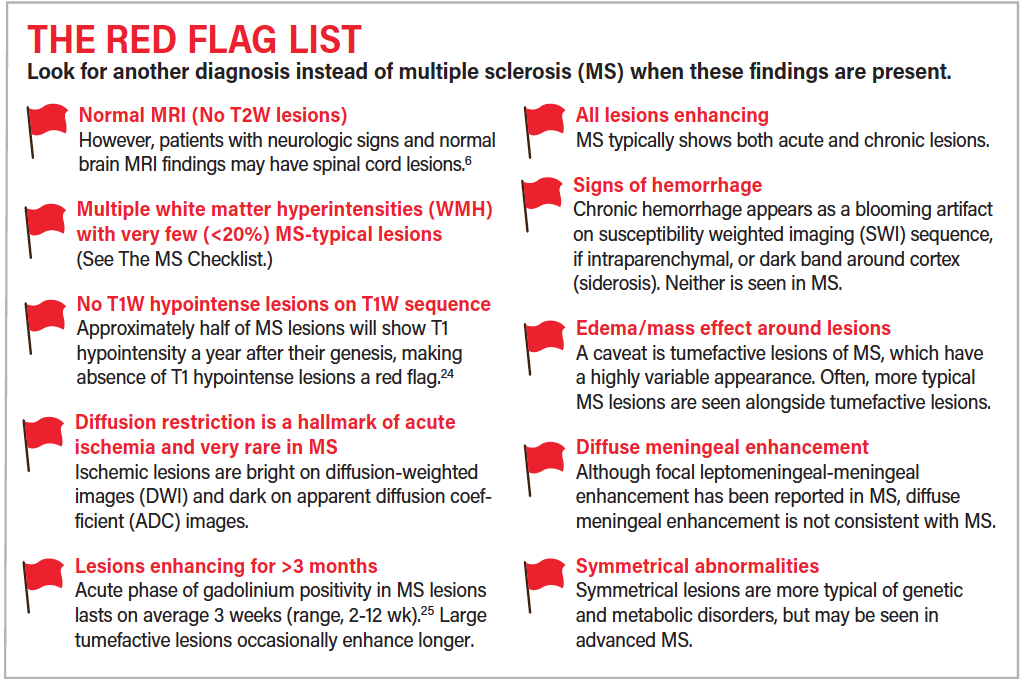
Look at the MRI for imaging red flags, like strokes, hemorrhages, cysts, findings that are too symmetric, subcortical, or normal. Remember that white matter lesions from migraine and microvascular disease are far more common that multiple sclerosis. NMO has differentiating features.
What does a bright spot on an MRI mean?
Bright spots on an MRI can develop due to conditions other than MS – including stroke, head trauma, migraine headache, or Vitamin B12 deficiency. Certain infections, or other autoimmune diseases such as lupus or sarcoidosis, are associated with increased lesions in the brain.
What causes an abnormal MRI?
Abnormal results may be due to: Abnormal blood vessels in the brain ( arteriovenous malformations of the head ) Tumor of the nerve that connects the ear to the brain ( acoustic neuroma ) Bleeding in the brain.
What does a dark spot on an MRI mean?
Definition. By Mayo Clinic Staff. A brain lesion is an abnormality seen on a brain-imaging test, such as magnetic resonance imaging (MRI) or computerized tomography (CT). On CT or MRI scans, brain lesions appear as dark or light spots that don’t look like normal brain tissue.
What color are lesions on MRI?
Sometimes MRI reports describe lesions as hyperintense, hypointense, or isointense. Hyperintense lesions are bright or white. In general, MS lesions are hyperintense or bright on T2 or FLAIR sequences. Hypointense lesions are dark or black.
How do I not worry about MRI results?
Try taking a walk. Regular exercise and a healthy diet have been shown to help alleviate stress and worry. If you find yourself feeling particularly worried about medical results, try taking a 30 minute walk. Or do another form of exercise that you enjoy.
How can an MRI tell if a tumor is cancerous?
Because of the different views, it’s possible to see where the tumor is, how big it is, and how it affects surrounding tissue structures. Magnetic resonance images can also show if a cancerous tumor has metastasized (spread) from its initial location to other parts of your body.
Does inflammation show on MRI?
MRI is an imaging method that is very sensitive in detecting inflammation and also bone erosions. This makes MRI an interesting tool to measure the course of the disease in randomised clinical trials and this suggests that MRI may also be useful in the diagnostic process.
What if an MRI shows something?
If you have a concern that your MRI revealed something that needs to be treated urgently, you can call your doctor’s office. However, if a radiologist identifies emergency findings, they will usually contact you. This is especially true if you require immediate treatment.
Can neurological damage be seen on MRI?
An MRI may be able help identify structural lesions that may be pressing against the nerve so the problem can be corrected before permanent nerve damage occurs. Nerve damage can usually be diagnosed based on a neurological examination and can be correlated by MRI scan findings.
Will a radiologist tell you if something is wrong?
“They aren’t doctors, and while they do know how to get around your anatomy, they aren’t qualified to diagnose you.” That is true even though the tech likely knows the answer to your question. Imaging techs administer thousands of scans a year.
How long does it take to get MRI results if something is wrong?
The Radiologist will send a report to the doctor who arranged the scan. They’ll discuss the results with you. It usually takes 1 to 2 weeks for the results of an MRI scan to come through, unless they’re needed urgently.
What does metastasis look like on MRI?
On MRI, metastases are usually iso- or hypointense on T1, hyperintense on T2, and exhibit avid enhancement [Figure 1]. Some metastases, such as melanoma, are T1 hyperintense due to the paramagnetic effects of melanin [Figure 3].
What is the most common cause of lesions in the brain?
Strokes are one of the most common causes of brain lesions, and you can often prevent a stroke, or at least delay when you have one or limit how severe it is.
What appears dark on T2 MRI?
CSF is dark on T1-weighted imaging and bright on T2-weighted imaging. A third commonly used sequence is the Fluid Attenuated Inversion Recovery (Flair).
What is the number 1 symptom of MS?
Numbness of the face, body, or extremities (arms and legs) is often the first symptom experienced by those eventually diagnosed as having MS.
What is the earliest symptom of MS?
Those symptoms include loss of vision in an eye, loss of power in an arm or leg or a rising sense of numbness in the legs. Other common symptoms associated with MS include spasms, fatigue, depression, incontinence issues, sexual dysfunction, and walking difficulties.
What shows up bright white on an MRI?
Axons are surrounded by a fatty material called myelin, which insulates them like a sheath and gives white matter its color. Abnormalities in white matter, known as lesions, are most often seen as bright areas or spots on MRI scans of the brain. They can reflect normal aging; white matter deteriorates as people age.
How many lesions does it take to be considered MS?
How long does it take to get MRI results if something is wrong?
The Radiologist will send a report to the doctor who arranged the scan. They’ll discuss the results with you. It usually takes 1 to 2 weeks for the results of an MRI scan to come through, unless they’re needed urgently.
Can MRI technician tell you results?
Legally, Medical Technologists Are Not Permitted Medical technologists are legally not allowed to discuss results with a patient. It doesn’t matter whether you are having an x-ray, ultrasound, computed tomography (CT), magnetic resonance imaging (MRI), or a nuclear medicine exam.
Should I be worried about my MRI?
An MRI is nothing to be nervous about. It will only give a better diagnosis and help your doctors provide the care you need. Call us today if you have any further questions about what to expect during your scan.